Home
/ Anatomy Of Musckes Sndctendons - Mr Miles Callahan Anatomy Of The Knee : Tendons connect the knee bones to the leg muscles that move the knee.
Anatomy Of Musckes Sndctendons - Mr Miles Callahan Anatomy Of The Knee : Tendons connect the knee bones to the leg muscles that move the knee.
Anatomy Of Musckes Sndctendons - Mr Miles Callahan Anatomy Of The Knee : Tendons connect the knee bones to the leg muscles that move the knee.. Anatomy describes isolated structures and also situates these structures within the larger functional systems. Muscles, either individually or in groups, are supported by fascia. New users enjoy 60% off. It specifically focuses on bones, muscles (including attachments, innervation, functions), arteries, veins, and nerves. Ebraheim's educational animated video describes the muscle anatomy of the hip and buttocks region with simple images;
On the other hand, the insertion is where a tendon attaches that muscle to the *more* movable bone. There are 10 intrinsic muscles located in the sole of the foot. In this lesson, we look at the muscle. Ebraheim's educational animated video describes the muscle anatomy of the hip and buttocks region with simple images; Lying exposed between the protective bones of the superiorly located ribs and the inferiorly located pelvic girdle, the muscles of this region play a critical role in protecting the.
Ligaments Tendons And Muscles Of The Hip Joint Naples Best Hip Surgeon from zehrcenter.b-cdn.net Ligaments connect two or more bones together and help stabilize joints. In this lesson, we look at the muscle. It is located toward the middle of the lower leg. New users enjoy 60% off. Lying exposed between the protective bones of the superiorly located ribs and the inferiorly located pelvic girdle, the muscles of this region play a critical role in protecting the. Tendons are the reason a muscle can move the bones in our body when muscles contract. Anatomy ankle anatomy ankle + ligament + tendon the foot anatomy human ankle anatomy 3d leg muscle lower leg anatomy leg articulation peroneal ankle muscles foot. The calf muscles (gastrocnemius and soleus), which are connected to the calcaneus via the achilles tendon.
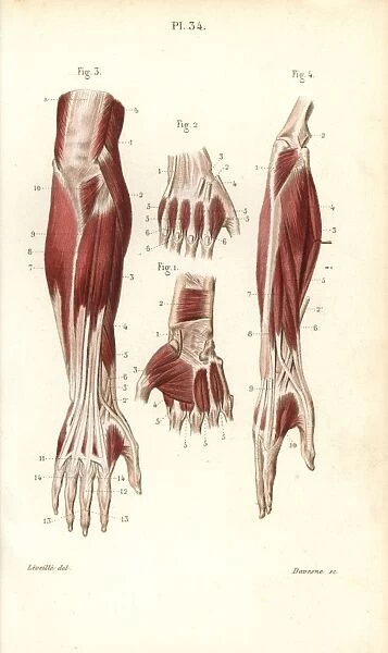
This is lesson 1 on the anatomy of the forearm.
Four muscles and their attached tendons make up the rotator cuff. Most structures in the foot are fairly superficial and can be easily palpated. The smaller bone that runs alongside the tibia (fibula) and the kneecap (patella) are the other bones that make the knee joint. The muscles of the abdomen, lower back, and pelvis are separated from those of the chest by the muscular wall of the diaphragm, the critical breathing muscle. Wrist anatomy is the study of the bones, ligaments and other structures in the wrist. The muscles of the plantar aspect are described in four layers. Tendons connect the knee bones to the leg muscles that move the knee. Lying exposed between the protective bones of the superiorly located ribs and the inferiorly located pelvic girdle, the muscles of this region play a critical role in protecting the. Anatomy of musckes sndctendons the foot is a part of vertebrate anatomy which serves the purpose of supporting the animal's weight and allowing for locomotion on land. When the muscle contracts, the tendons are pulled, and the bone is moved. They act collectively to stabilise the arches of the foot, and individually to control movement of the digits. There are two sets of flexor foot tendons, each made up of two muscles and tendons, one set that bends the big toe, the other set that bends the remaining four toes. Similar to ligaments, they are made of collagen and can withstand increased tension.
See tendons muscles foot lower leg anatomy stock video clips. The majority of muscles in the leg are considered long muscles, in that they stretch great distances. Tendons attach muscle to bone. In this lesson, we look at the muscle. Related posts of diagram of shoulder muscles and tendons muscle anatomy coloring sheets.
What Is A Tendon Anatomy Definition Video Lesson Transcript Study Com from study.com However, it is susceptible to injury, especially from repetitive strain. As these muscles contract and relax, they move skeletal bones to create movement of the body. Tendons connect the knee bones to the leg muscles that move the knee. The muscles of the abdomen, lower back, and pelvis are separated from those of the chest by the muscular wall of the diaphragm, the critical breathing muscle. Similar to ligaments, they are made of collagen and can withstand increased tension. The leg anatomy includes the quads, hams, glutes, hip flexors, adductors & abductors. There are two sets of flexor foot tendons, each made up of two muscles and tendons, one set that bends the big toe, the other set that bends the remaining four toes. The calf muscles (gastrocnemius and soleus), which are connected to the calcaneus via the achilles tendon.
New users enjoy 60% off.
Tendons and ligaments are bands of connective tissue that help stabilize the body and allow movement. The lower leg is also home to nerve fibers. Four muscles and their attached tendons make up the rotator cuff. A tendon connects the muscle to the bone. Tendons vary in size and are somewhat elastic and attach bones to muscles. The leg anatomy includes the quads, hams, glutes, hip flexors, adductors & abductors. The peroneal muscles (peroneus longus and peroneus brevis), on the outside edge of the ankle and foot. Anatomy describes isolated structures and also situates these structures within the larger functional systems. Anatomy is a road map. The fibula, or calf bone, is smaller and is located on the outside of the lower leg. Muscles and tendons tendons are thick bands of connective tissue that connect muscle to bone. Muscle anatomy coloring sheets 12 photos of the muscle anatomy coloring sheets free muscle anatomy coloring sheets, muscle anatomy coloring pages, muscle anatomy coloring pages free, muscle anatomy coloring sheets, human muscles, free muscle anatomy coloring sheets, muscle anatomy coloring pages. Anatomy ankle anatomy ankle + ligament + tendon the foot anatomy human ankle anatomy 3d leg muscle lower leg anatomy leg articulation peroneal ankle muscles foot.
A solid understanding of anatomy is essential to effectively diagnose and treat patients with foot and ankle problems. Tendons attach muscle to bone. They act collectively to stabilise the arches of the foot, and individually to control movement of the digits. The majority of muscles in the leg are considered long muscles, in that they stretch great distances. There are 10 intrinsic muscles located in the sole of the foot.
Muscles And Tendons Of The Forearm And Hand Print 14216190 from www.prints-online.com When the muscle contracts, the tendons are pulled, and the bone is moved. Most structures in the foot are fairly superficial and can be easily palpated. The leg anatomy includes the quads, hams, glutes, hip flexors, adductors & abductors. The calf muscles (gastrocnemius and soleus), which are connected to the calcaneus via the achilles tendon. The quadriceps muscles provide strength and power with knee extension (straightening). Included are several layered views of the back muscles, the dorsal muscles, subclavius muscles, rhomboideus major and minor muscles, deltoid muscles and many more. There are numerous tendons around the knee that also help to stabilize the knee. Ebraheim's educational animated video describes the muscle anatomy of the hip and buttocks region with simple images;
These muscles allow the ankle to bend downward and outward.
On the other hand, the insertion is where a tendon attaches that muscle to the *more* movable bone. This anatomy chart is a great example of beauty and function in one, as it is pleasing to look… Muscles and tendons tendons are thick bands of connective tissue that connect muscle to bone. In this lesson, we look at the muscle. Tendons and ligaments are bands of connective tissue that help stabilize the body and allow movement. The majority of muscles in the leg are considered long muscles, in that they stretch great distances. Anatomy describes isolated structures and also situates these structures within the larger functional systems. When muscles contract, they pull on the tendons to move the bones. Lesson on the anatomy of the forearm: This is lesson 1 on the anatomy of the forearm. They are associated with muscles discussed in the section above (see above). Anatomy ankle anatomy ankle + ligament + tendon the foot anatomy human ankle anatomy 3d leg muscle lower leg anatomy leg articulation peroneal ankle muscles foot. Related posts of diagram of shoulder muscles and tendons muscle anatomy coloring sheets.

